 |
History of the VOXEL-MAN Project at the University Medical Center |
de | en |
| x | 1969 First Computer at the UKE
Prof. Klaus Dieter Vogt (1921-2004), head of the Clinical Chemical Laboratory and Dr. Hans Otto Wüster (1927-1985), head of Information Technology at the Deutsches Elektronen-Synchrotron DESY in Hamburg agree on a cooperative study for investigating the potential of computer application in medicine. DESY sets up the first computer in the medical center for this project. The physicist Dr. Karl Heinz Höhne (*1937) and coworkers develop a system for acquisition and analysis of laboratory tests. With the system ("LABMAT", in routine use until 1974), one of the first worldwide, it is shown that the application of computers decisively improves efficiency and quality of a clinical laboratory.
1971 Creating the First Digital Images: Computer Analysis of Scintigrams
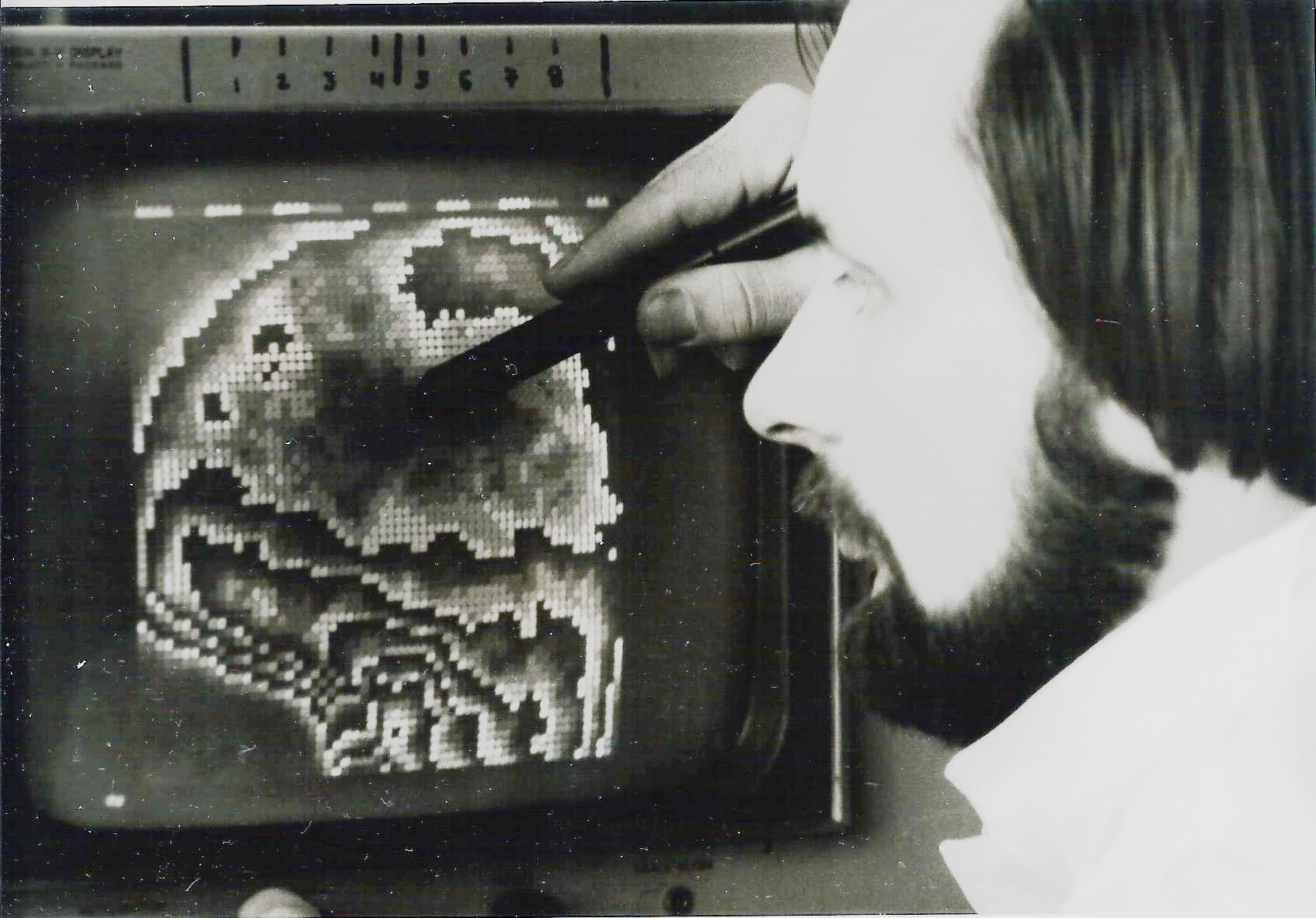
On the initiative of Dr. Dragutin Nowak the DESY working group develops together with Prof. Claus Schneider and Dr. Ricardo Montz (Department of Nuclear Medicine) a novel system (system ISAAC) for interactive analysis of images indicating the distribution of radioactivity in the body delivered by a gamma camera (scintigrams). Digital images are at their infancy at that time, the term "pixel" was only known to a handful of experts. For the analysis of the scintigrams one of the first bitmap displays (64X64 pixels, 16 gray levels) is developed within the project (today one would call this a graphics card). The interaction with the system is via a "light pen". One of the successful applications is the detection of brain death. ISAAC is in routine use for 5 years until its replacement by a commercial system.
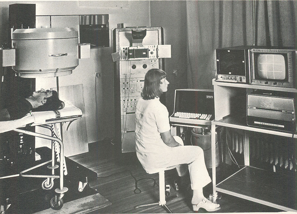
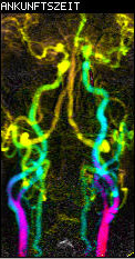
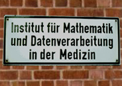
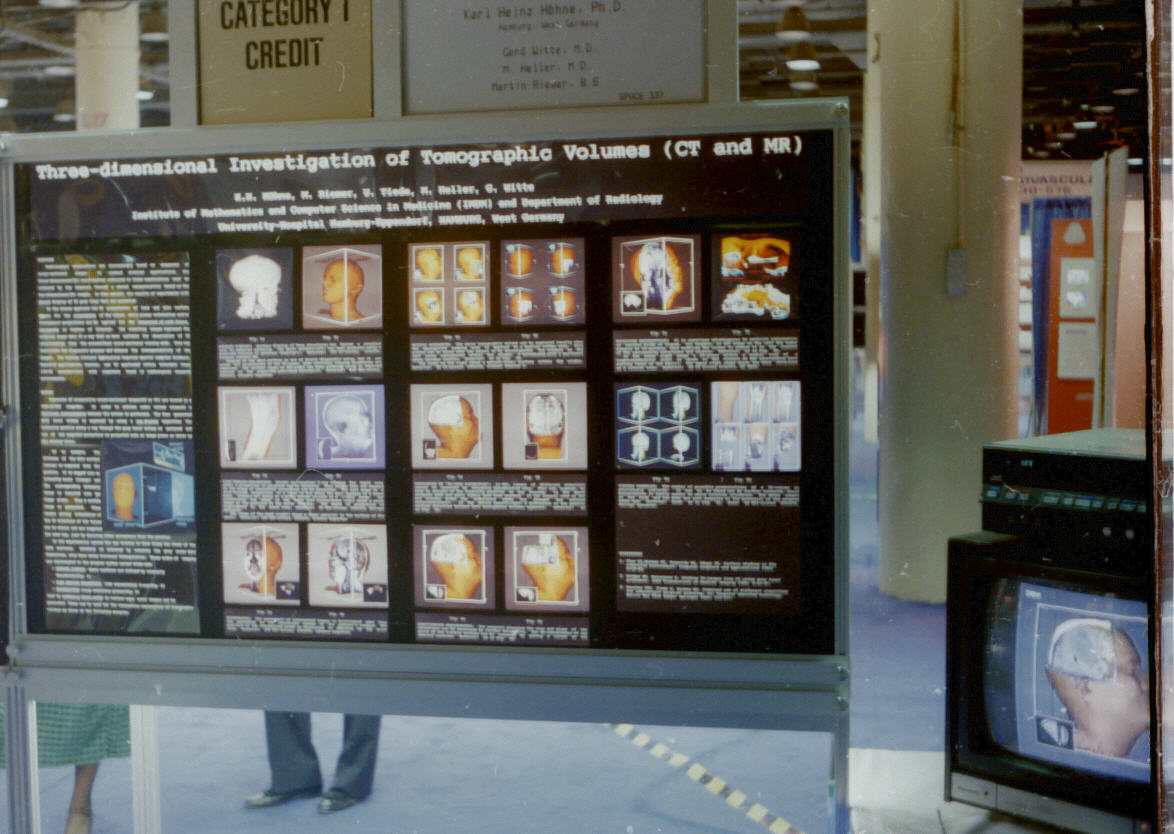
The DESY working group develops in cooperation with the Department of Radiology (Prof. Egon Bücheler) a system for quantitative analysis of angiograms, sequences of X-ray images showing the propagation of a contast medium. The objective is the diagnostic quantification of blood perfusion in organs such as the kidney, liver, lung and the heart. The project is funded by the government with one million DM. Cutting edge computer science tools are developed for digital image sequence processing: A multiprocessor system for real time acquisition and display of image sequences, special computer languages for their analysis and storage in a data base. The latter one runs on a mainframe computer at DESY connected with a 40,8 kbits/s data line. However, the project does not meet the expectations for diagnostic radiology. 1978 Institute of Mathematics and Computer Science in Medicine
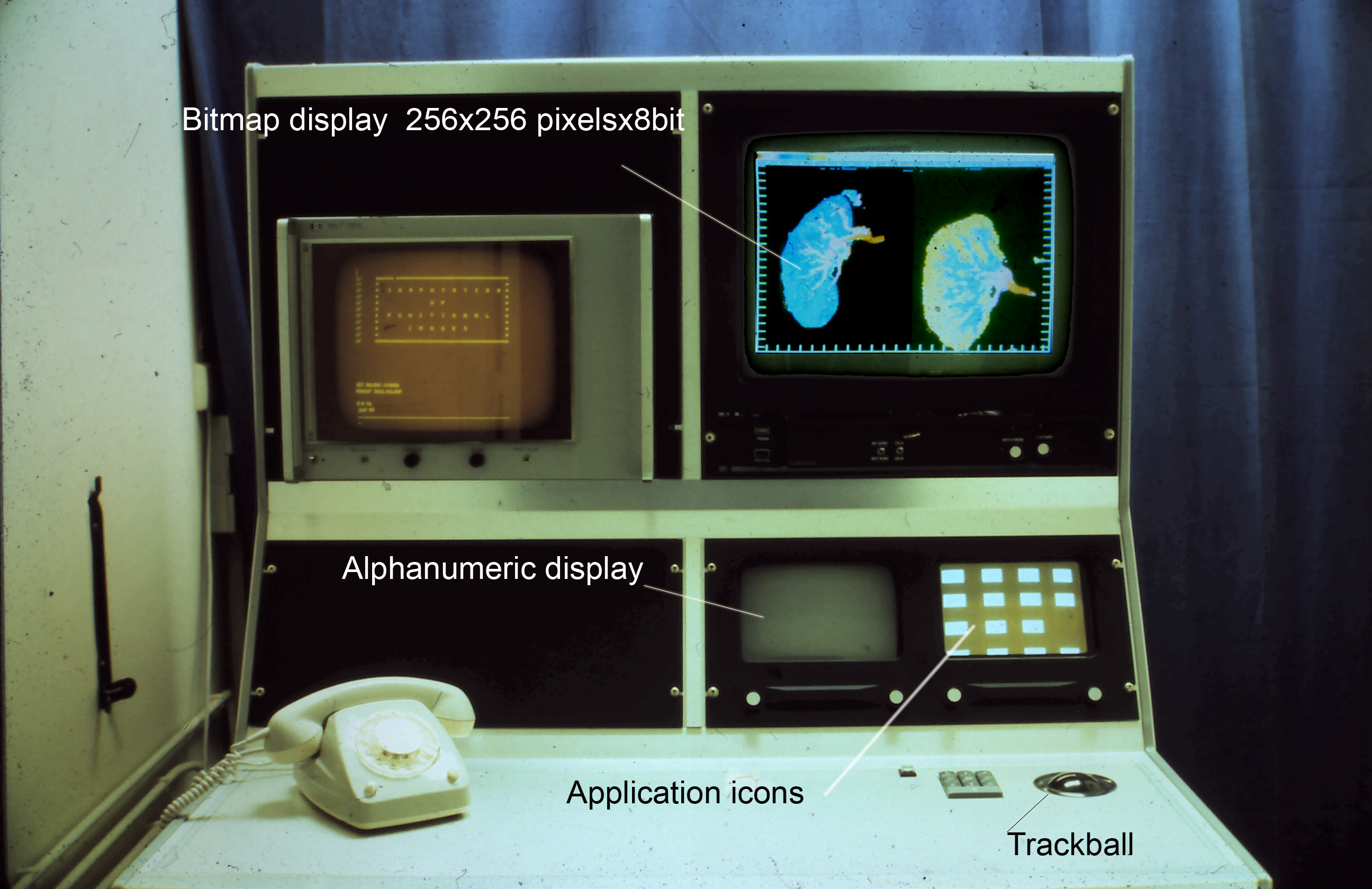
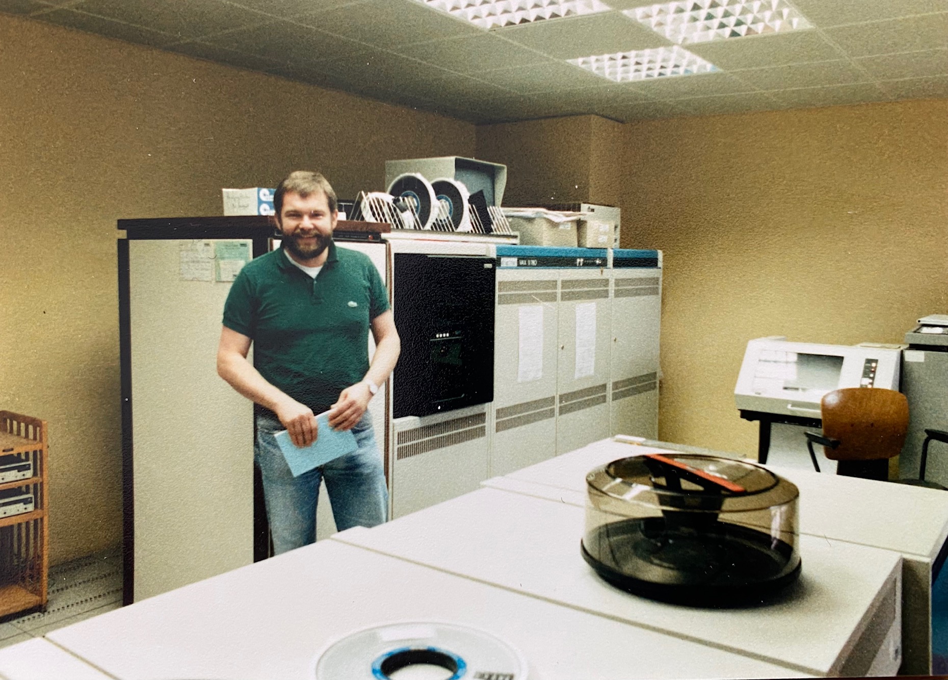
Karl Heinz Höhne is appointed professor and director of the Department of Computer Science in Medicine within the newly founded institute. The department gets a VAX-11/780 computer, which was extremely powerful for the time (4 Mb, later 32 Mb main memory, tape and removable disk drives, total costs including conversions approx. 1.5 million DM). The joint UKE-DESY working group that has now emerged becomes even more powerful, can turn to new projects such as image databases and 3D reconstruction of tomographic image sequences.
1978-1985 Development of Concepts for Picture Archiving and Communication Systems (PACS)
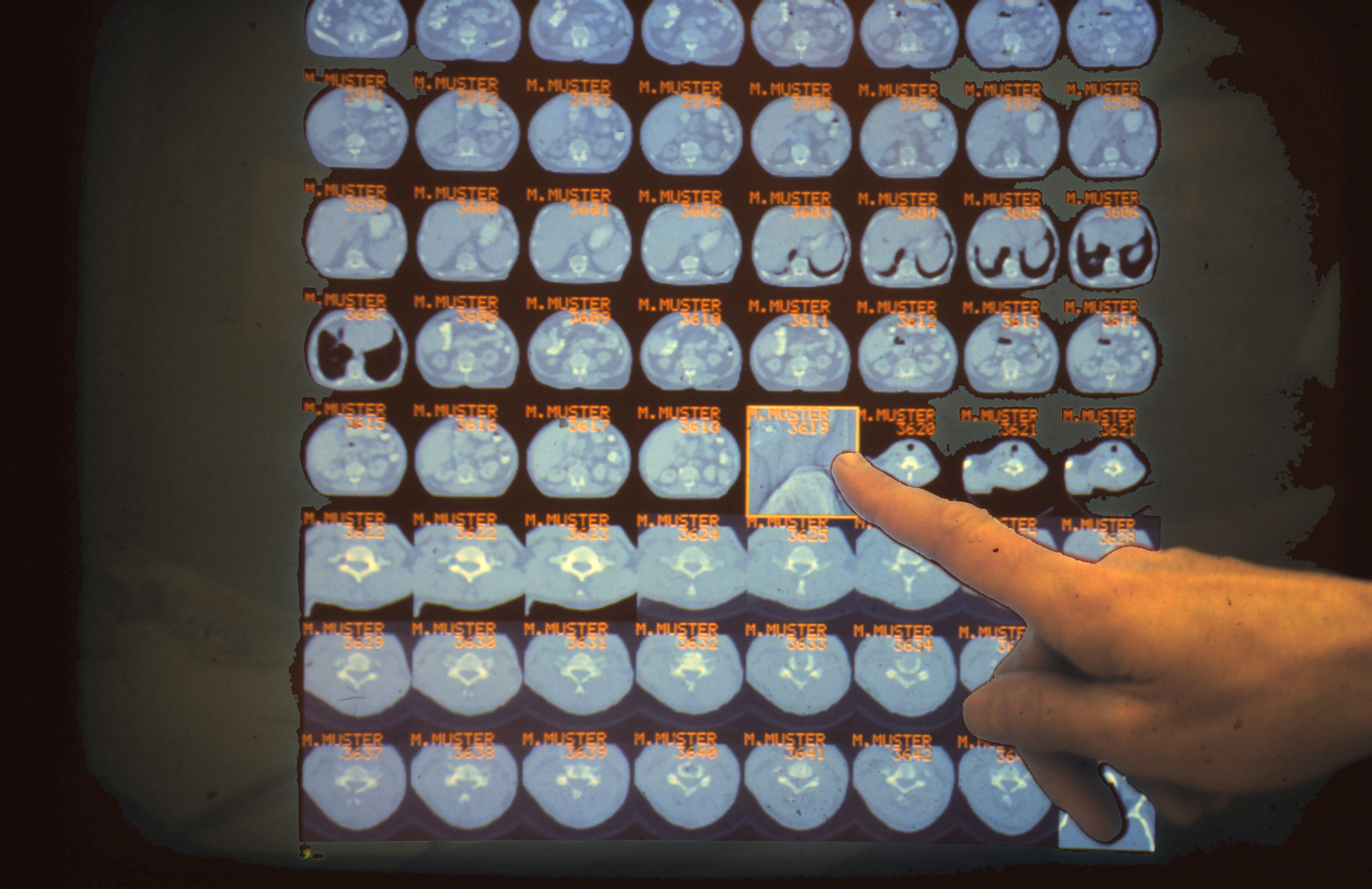
Digital images become more and more common in diagnostic radiology. Using a specially developed language (ISQL - image SQL) a user interface is created for an Oracle data base containing the image data. A demonstration data base, already then with a touch screen, shows the feasibility of the concept. For the discussion of the state of the art the institute organizes in 1984 the NATO Advanced Study Institute "Pictorial Information Systems in Medicine" in Braunlage, Germany, where EUROPACS, the European Association for Picture Archiving and Communication Systems (PACS) is founded. Because of lacking network and hardware standards and capabilities the method is not mature for a broad application at that time. Since the challenges are more organizational than scientific, PACS research is no longer pursued. 1983 First 3D Reconstructions from Microscopic Specimens
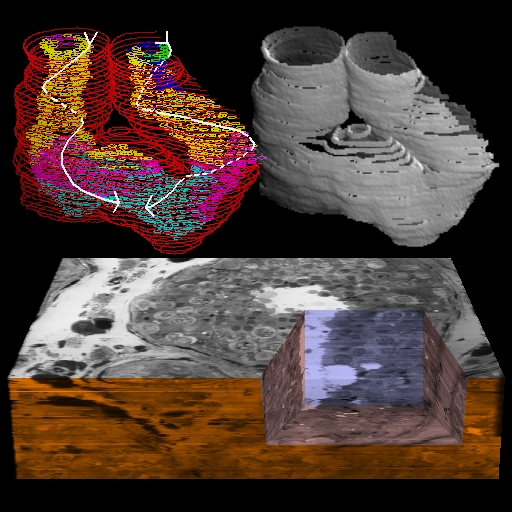
In cooperation with the Department of Microscopic Anatomy (Prof. Adolf Friedrich Holstein and Dr. Wolfgang Schulze) for the first time 3D views of the human seminiferous tubules are computed from microscopic image sequences. It is shown that the production of sperms is located on a spiral according to their state of maturity.
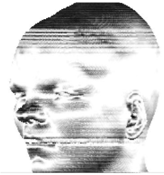
1984 First Reconstructions from Radiological Images Inspired by the early work of Gabor Herman (Philadelphia) in cooperation with the Department of Radiology first reconstructions from computer tomograms (CT) and magnetic resonance tomograms (MRI) are created. They are shown at the Congress of the German Radiological Society in 1984 and more or less received as gimmicks.
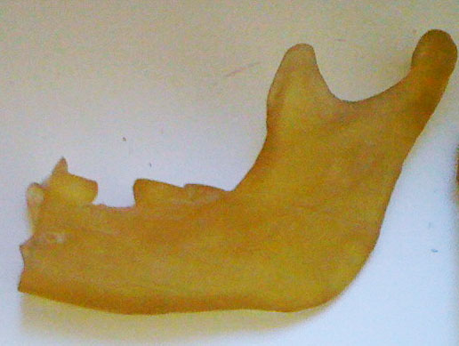
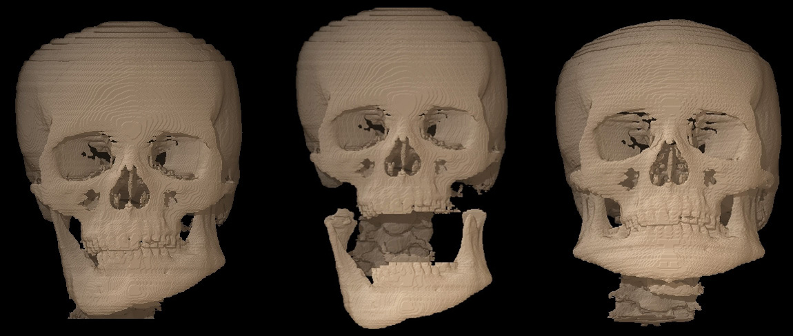
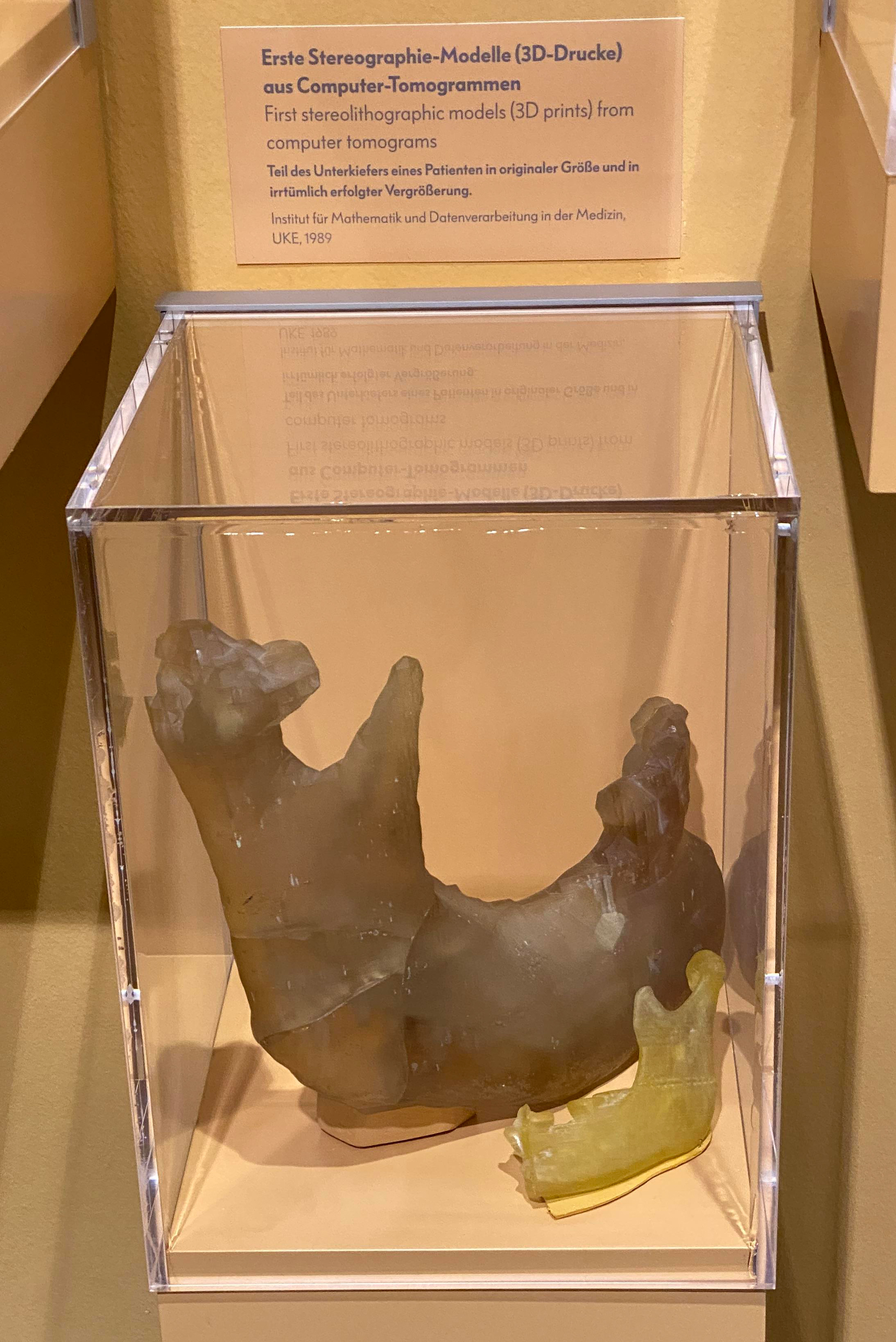
Prof. Hans-Joachim Höltje of the Department of Dental, Oral and Maxillo-facial Surgery of the UKE learns at a congress in Philadelphia about the developments in his own Medical School. From the first contact a successful cooperation evolves lasting for years. Among the first in the world 3D models (also as 3D prints by stereolithography) are used at the UKE for planning craniofacial interventions in clinical routine.
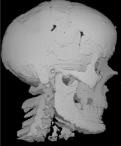
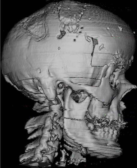
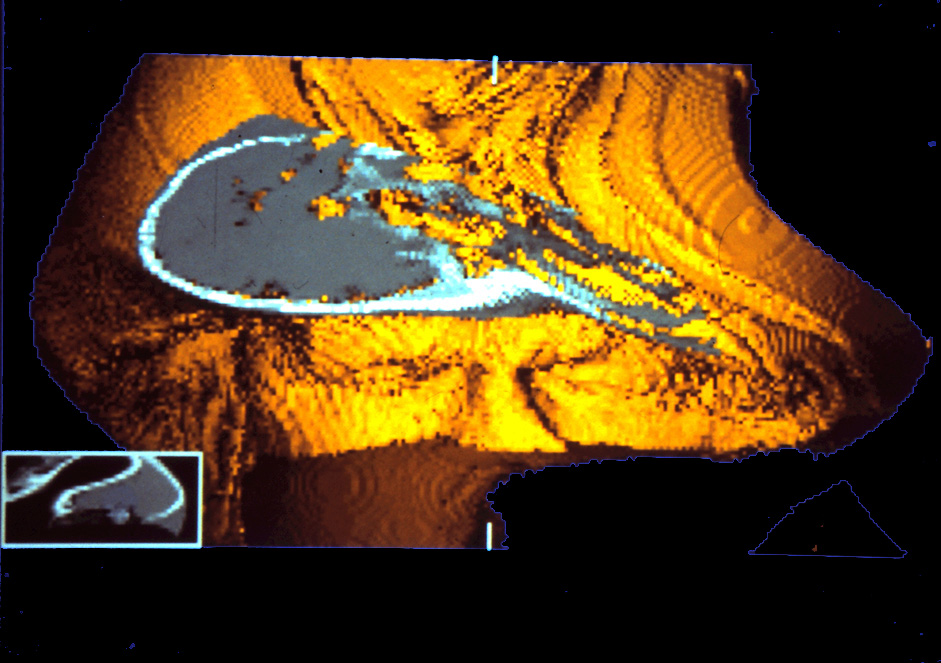
During a sabbatical at the IBM Palo Alto Scientific Center in the Silicon Valley Karl Heinz Höhne has access to computers (IBM 3081) with a capacity exceeding those available in Hamburg by far. Using them he pioneers together with Ralph Bernstein a novel graphics algorithm ("gray scale gradient shading ") that creates 3D views with unprecedented realism*. This method becomes the standard for the 3D visualization from Computer Tomography (CT) and Magnetic Resonance Imaging (MRI). While the computation of a single view takes minutes in 1985, today (2016) a modern PC can compute them in real time. * Karl Heinz Höhne, Ralph Bernstein: Shading 3D-images from CT using gray level gradients . IEEE Trans. Med. Imaging 5, 1 (1986), 45-47.
1986-1988 A Series of Innovations The novel method is tested with image data delivered by the Department of Radiology and Siemens. For the first time it allows the 3D view to the brain (1986,1987) and the beating heart (1988) of living persons via Magnetic Resonance Imaging (MRI). The demonstrations, partly already in stereoscopic mode, are highlights at the congresses of the Radiological Society of North America (RSNA) in Chicago in these years. The Institute of Mathematics and Computer Science in Medicine becomes a destination of visitors from all over the world.
1986 The Project is Given the Name "VOXEL-MAN" The name is derived from the term Voxel, the description of a three-dimensional pixel.The 3D models of the VOXEL-MAN Project are compounds of such voxels. Occasionally the name Voxel-Man is also used as a general term for a digital representation of the human body.
The expectation that the new possibilities would be soon accepted as instruments in radiology or surgical disciplines such as neurosurgery does not become true. It turns out that 3D imaging is not a substantial advance in radiological diagnosis. For surgical planning and simulation the advance is obvious. However, the step into the virtual world is too big, the more so as with the then available hardware the handling of the new tools is time-consuming and cumbersome. Today preoperative and even intraoperative 3D visualization is standard in many surgical disciplines.
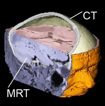
While in Computer Tomography (CT) dense objects (bone, metal) show up very well, Magnetic Resonance Tomography (MRT) is better in depicting soft tissue. A method for combining the two modalities in a single 3D model is developed.
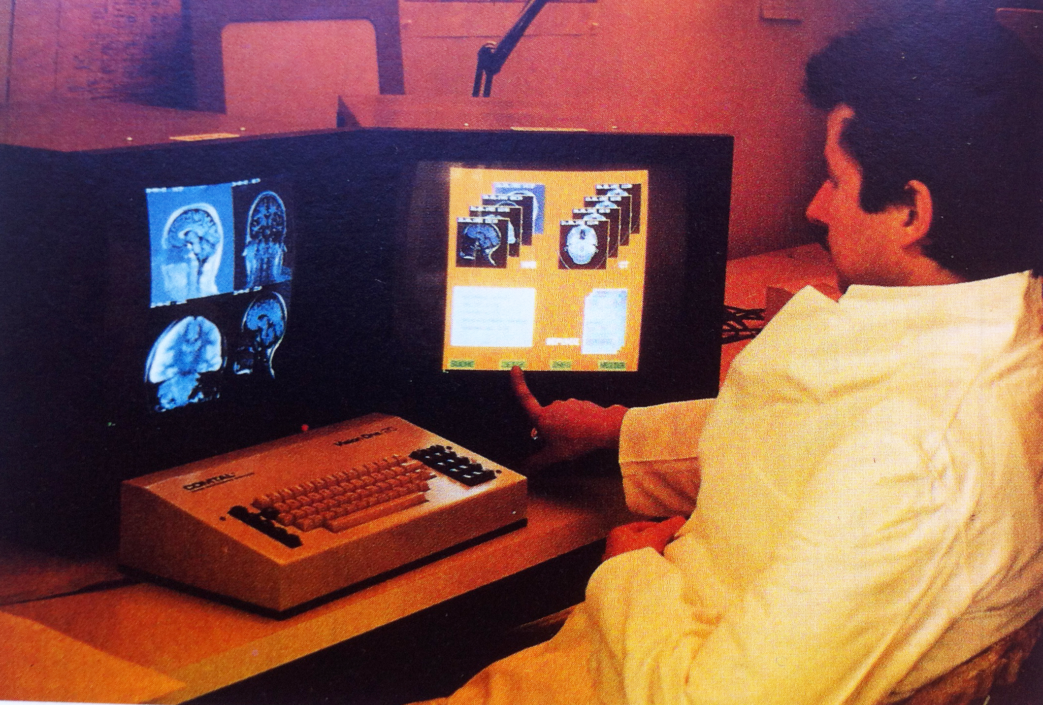
1990 VOXEL-MAN as Add-on in the Siemens Magnetom Tomograph As first commercial device at all the Siemens Magnetom tomograph offers an early version of the VOXEL-MAN software for 3D visualization. Yet because of the limited computing capacity of computers at that time (the computation of a single view takes several minutes) it does not find a broad acceptance. Today (2016) such program is part of any Computer or Magnetic Resonance Tomograph. 1990 Prof. Lierse is excited about VOXEL-MAN
Because of the low acceptance in clinical medicine the researchers change their focus to computer based 3D models for education and training. In cooperation with the neuroanatomist Prof. Werner Lierse (1928-1993) novel method of combination 3D models with anatomical and radiological knowledge is developed. For the project Prof. Lierse spends more time in the Institute of Mathematics and Computer Science in Medicine than in his own institute.
1991 Linking Models with Descriptive Knowledge
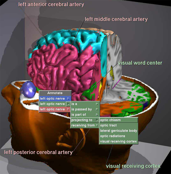
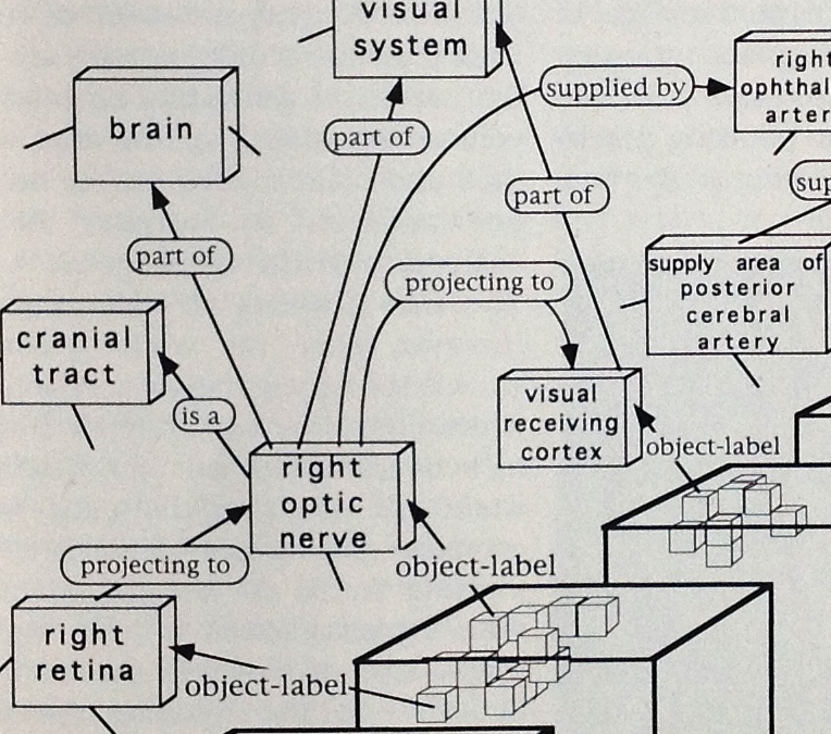
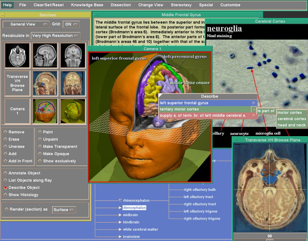
The necessity of linking descriptions to the 3D models leads involuntarily to dealing with methods of knowledge representation. Based on the method of "semantic networks" a concept is developed that allows such linking of knowledge about organs to the image data. As a result a user can navigate in the descriptions and ask for the anatomical correlate to be marked or he can vice versa point on an area in the 3D model for inquiring information e. g. about name and relation to other organs, function, or supplying blood vessel.
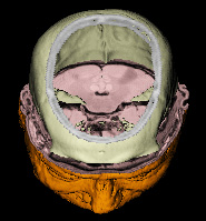
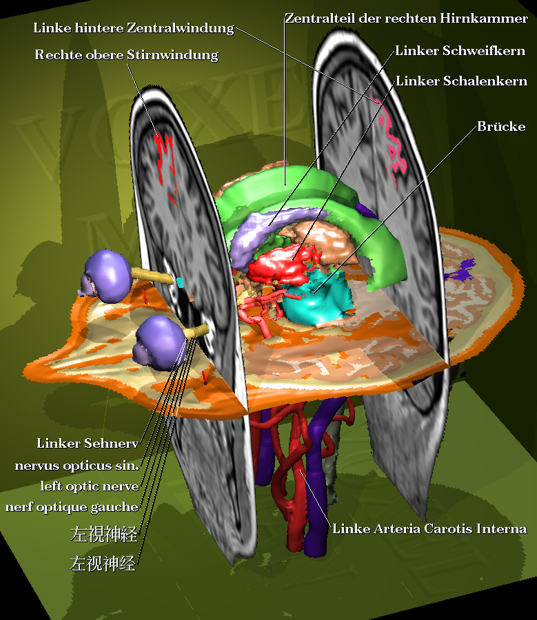
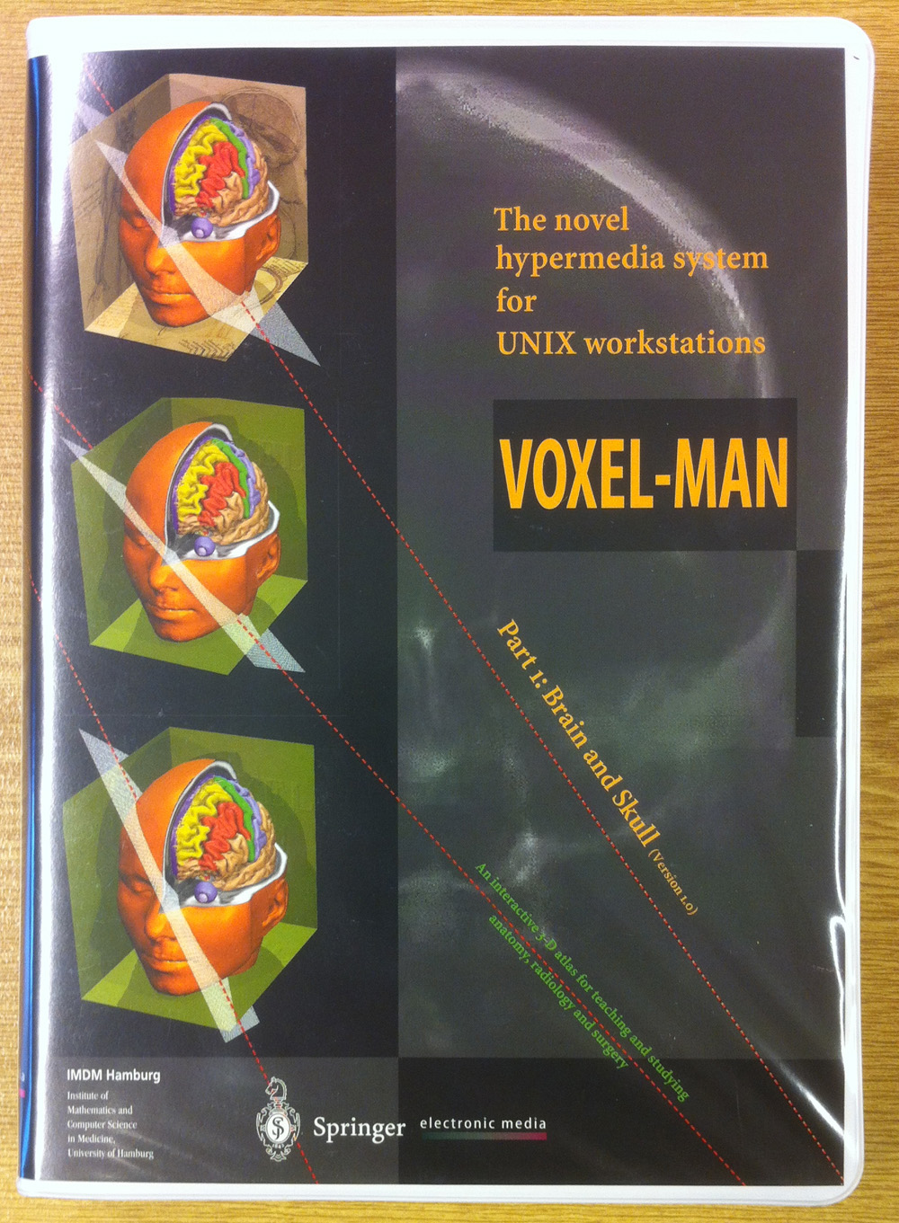
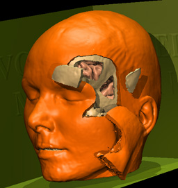
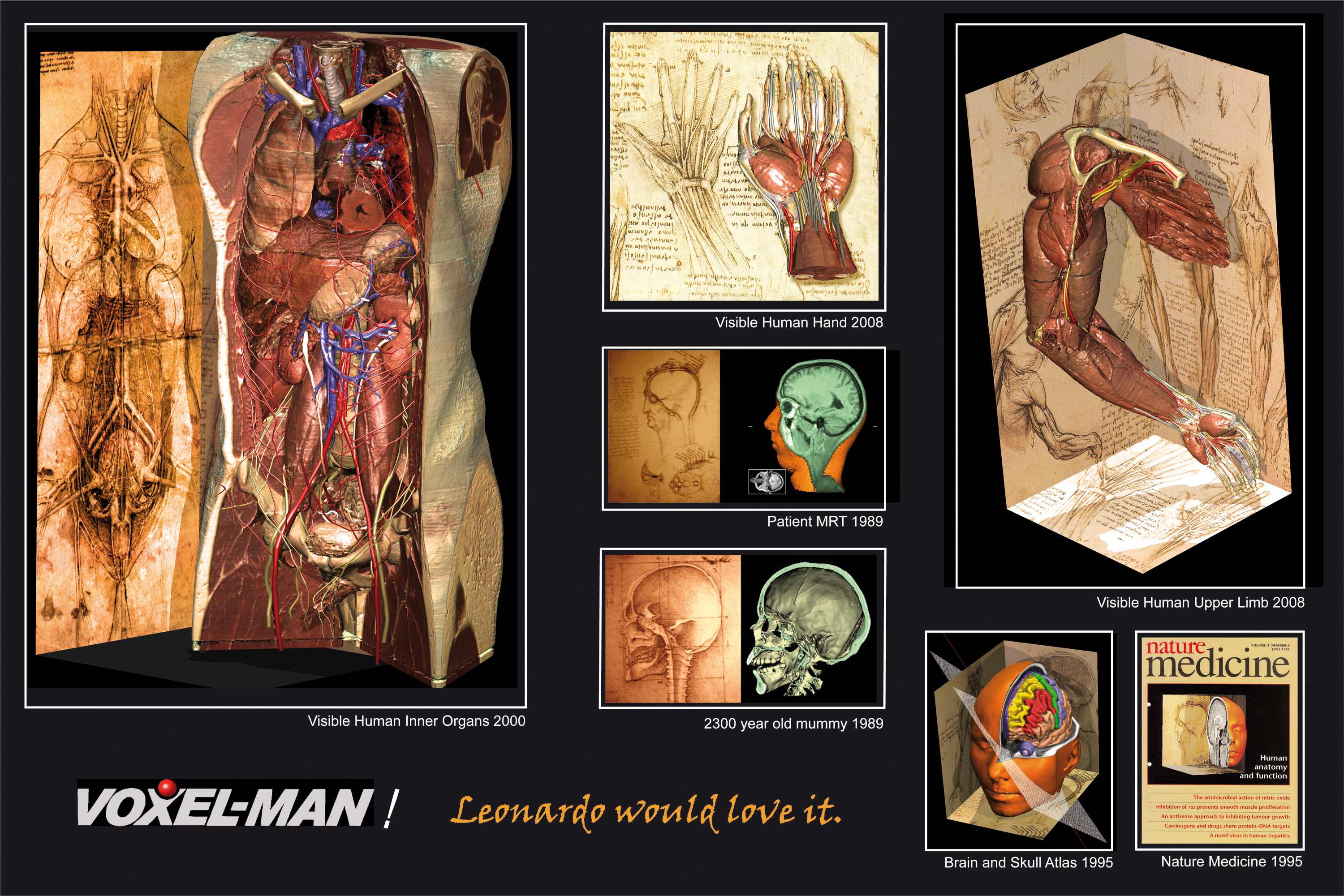
Based on this concept the first interactive 3D atlas of the human brain and skull is created. Unexpectedly Prof. Lierse passes away in 1993 and cannot experience its final version. It is completed under the supervision of Prof. Udo Schumacher (Institute of Anatomy and Experimental Morphology). The atlas consists of a 3D computer model of the human brain containing 250 anatomical objects. At the computer screen they can be viewed from any direction, dissected, disassembled, interrogated or shown in their radiological manifestation. Annotations can be chosen in five different languages (German, English, French, Japanese and Chinese). Commentary of the American Journal of Neuroradiology* about visitors experiencing the atlas at the RSNA Congress in Chicago 1991: ".................they were looking into the future." In 1995 it is published by Springer. The downsized version VOXEL-MAN 3D Navigator can be downloaded here. * Am. J. Neuroradiology 14, 3 (1993), 560
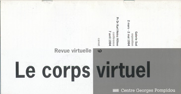
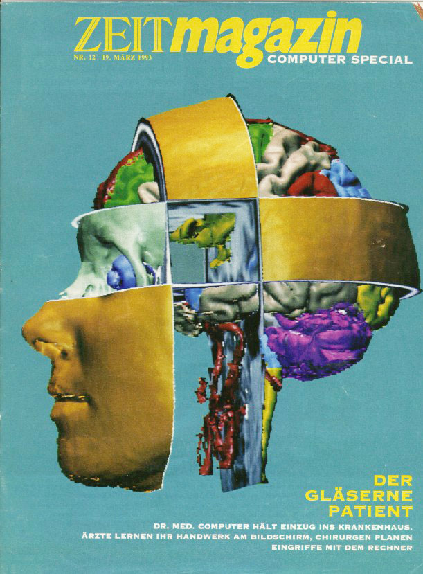
It turns out that the novel images are an attraction beyond medicine. So the animations of the VOXEL-MAN Project are in the center of the exhibition "Le Corps Virtuel" at the Centre Pompidou in Paris. The newspaper "Le Monde"*: "..........fort spectaculaire (definitely spectacular)". * Le Monde, 7. April 1994
1994 Virtual Endoscopy
From the beginning the VOXEL-MAN software allows a perspective view from every point of view, including from inside the body. Yet altough the technique is available, others have the idea earlier to use it for the simulation of endoscopic procedures ("Virtual Endoscopy").
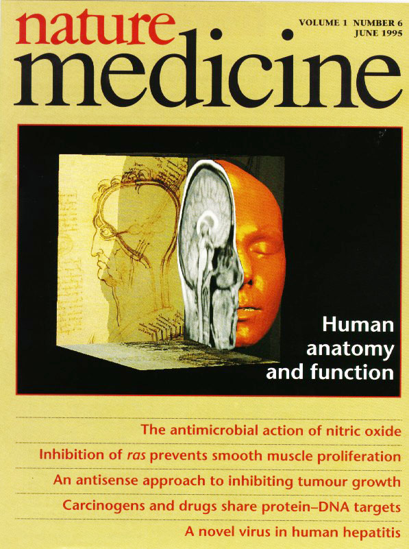
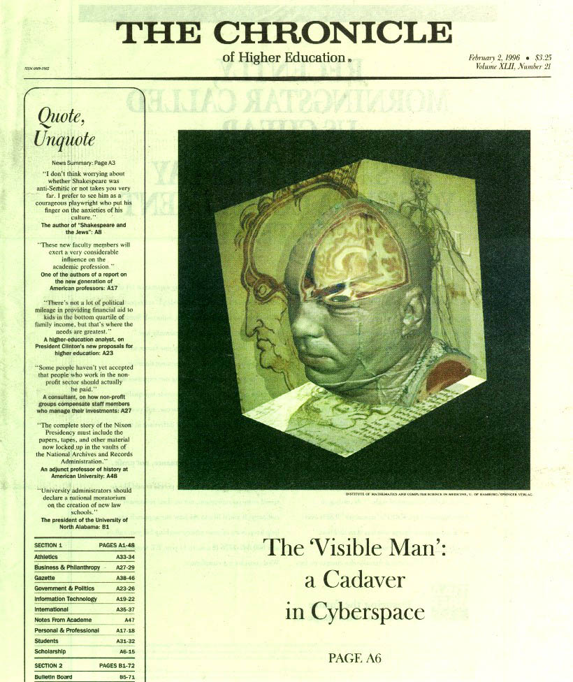
1995 VOXEL-MAN on the cover of the Journal Nature Medicine
At the occasion of the publication A new representation of knowledge concerning human anatomy and function* a composition created with the VOXEL-MAN system appears on the cover of Nature Medicine, which confronts computer-generated anatomy with a drawing by Leonardo da Vinci. * Nat. Med. 1, 6 (1995), 506-511
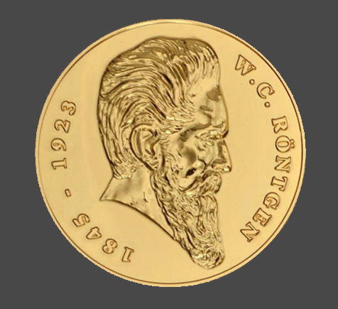
1996 Roentgen Medal
Karl Heinz Höhne receives the Roentgen Medal for "Fundamental work on 3D visualization of realistic anatomy from CT and MRI image data".
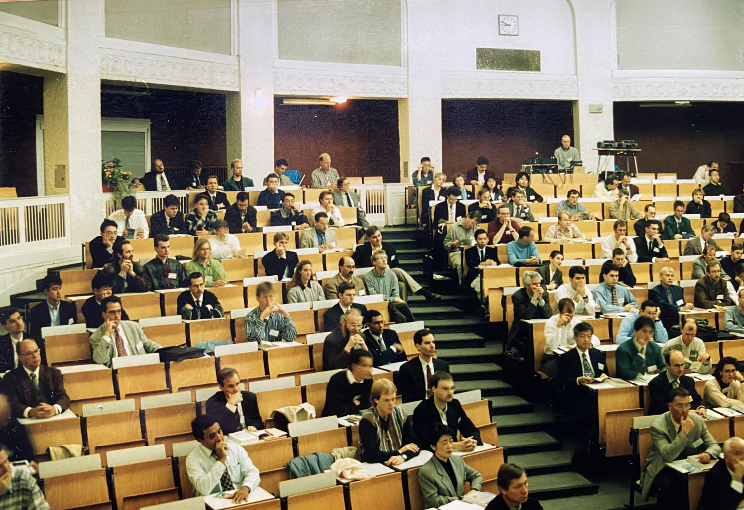
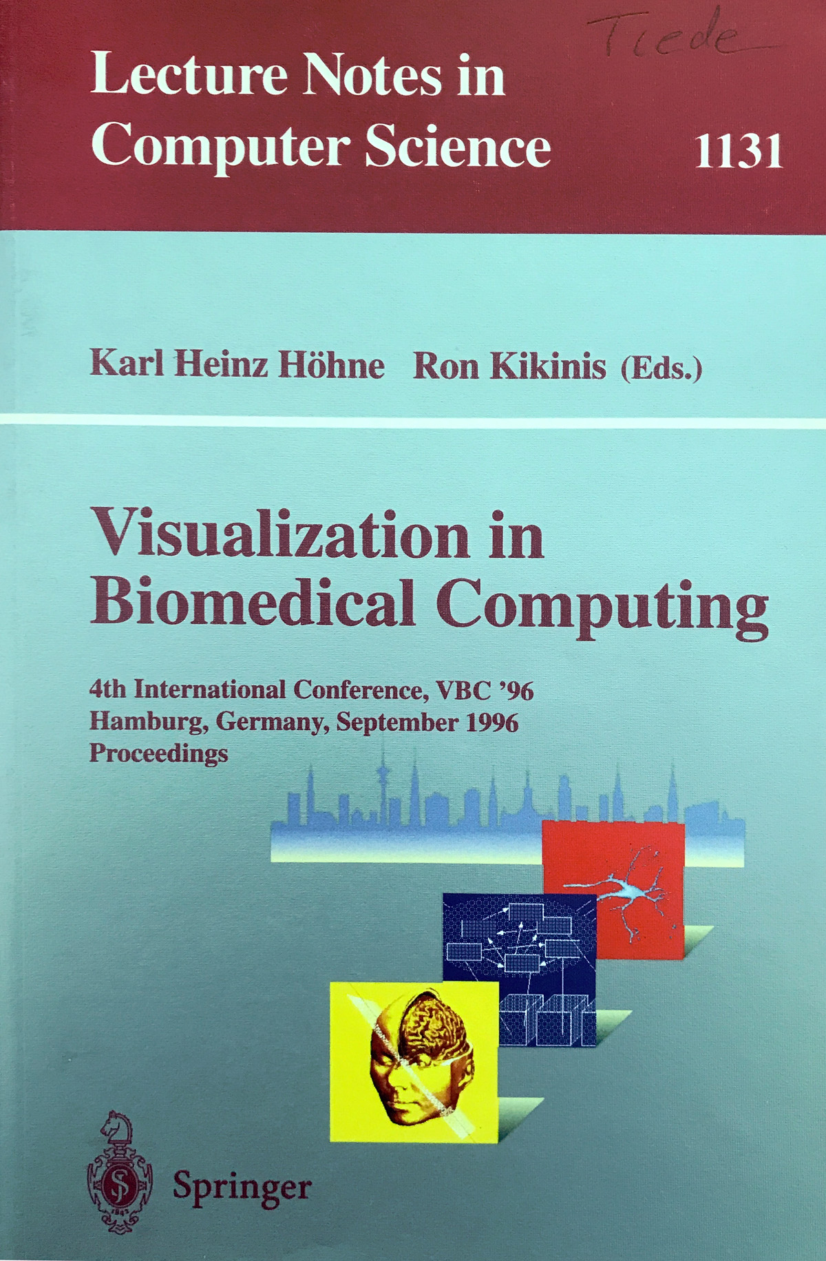
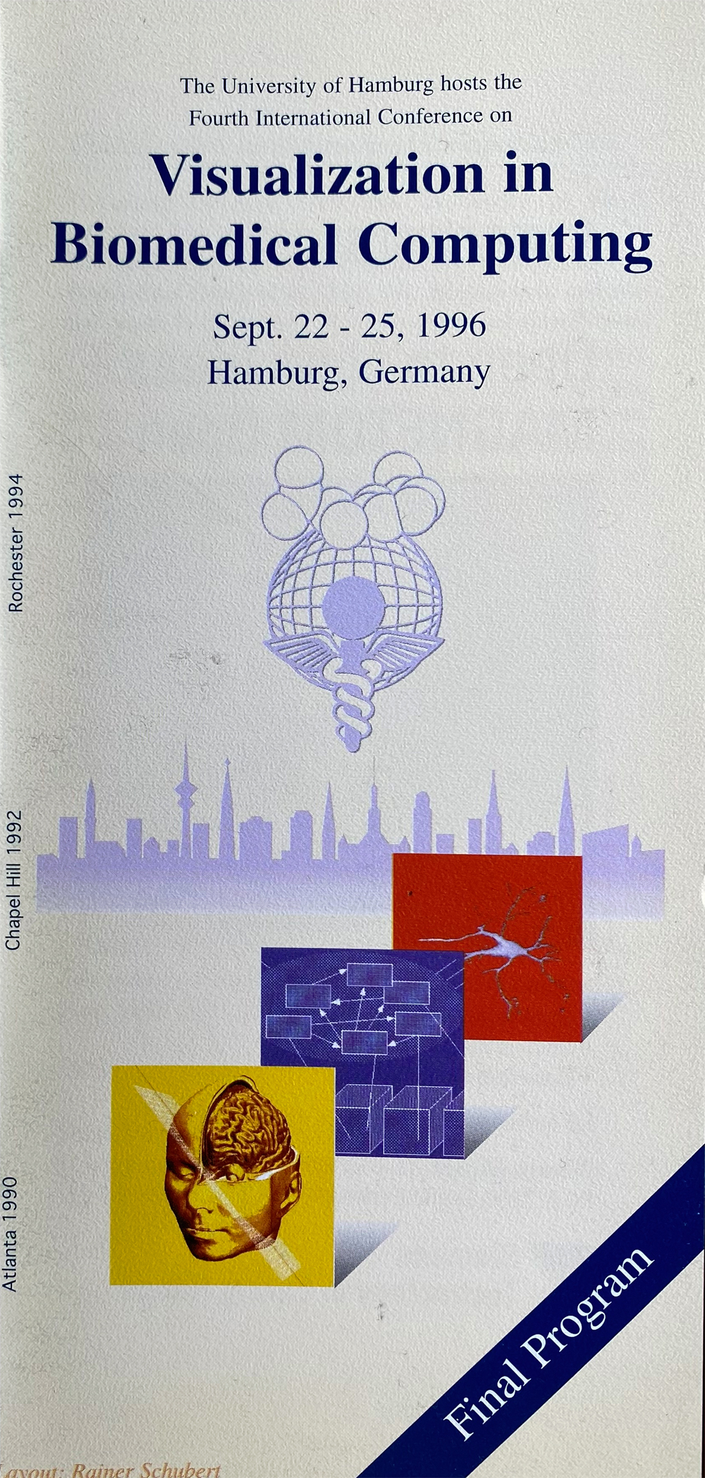
1996 Visualization in Biomedical Computing in Hamburg
In 1996 the IMDM runs the 4th Conference on Visualization in Biomedical Computing with about 220 participants. At this occasion the merger with the conferences CVRMed (Computer Vision, Virtual Reality and Robotics in Medicine) and MRCAS (Medical Robotics and Computer Assisted Surgery) into the conference and later the Society for Medical Image Computing and Computer Assisted Intervention (MICCAI) was decided.
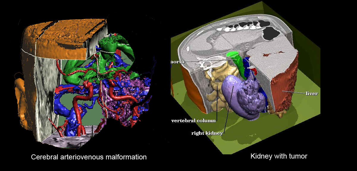
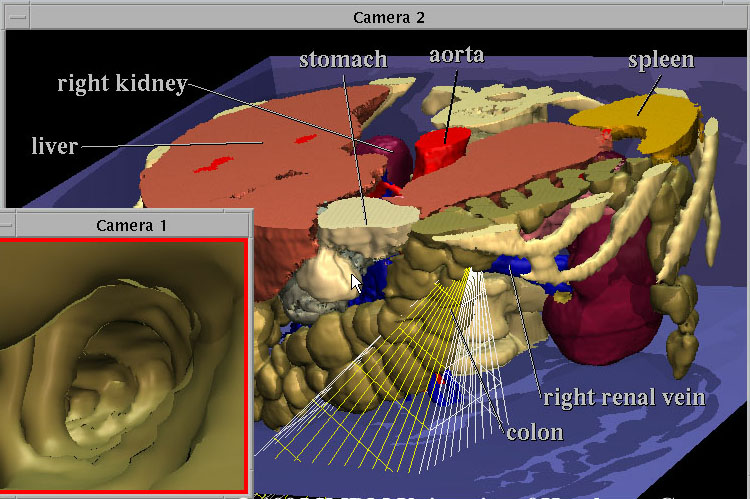
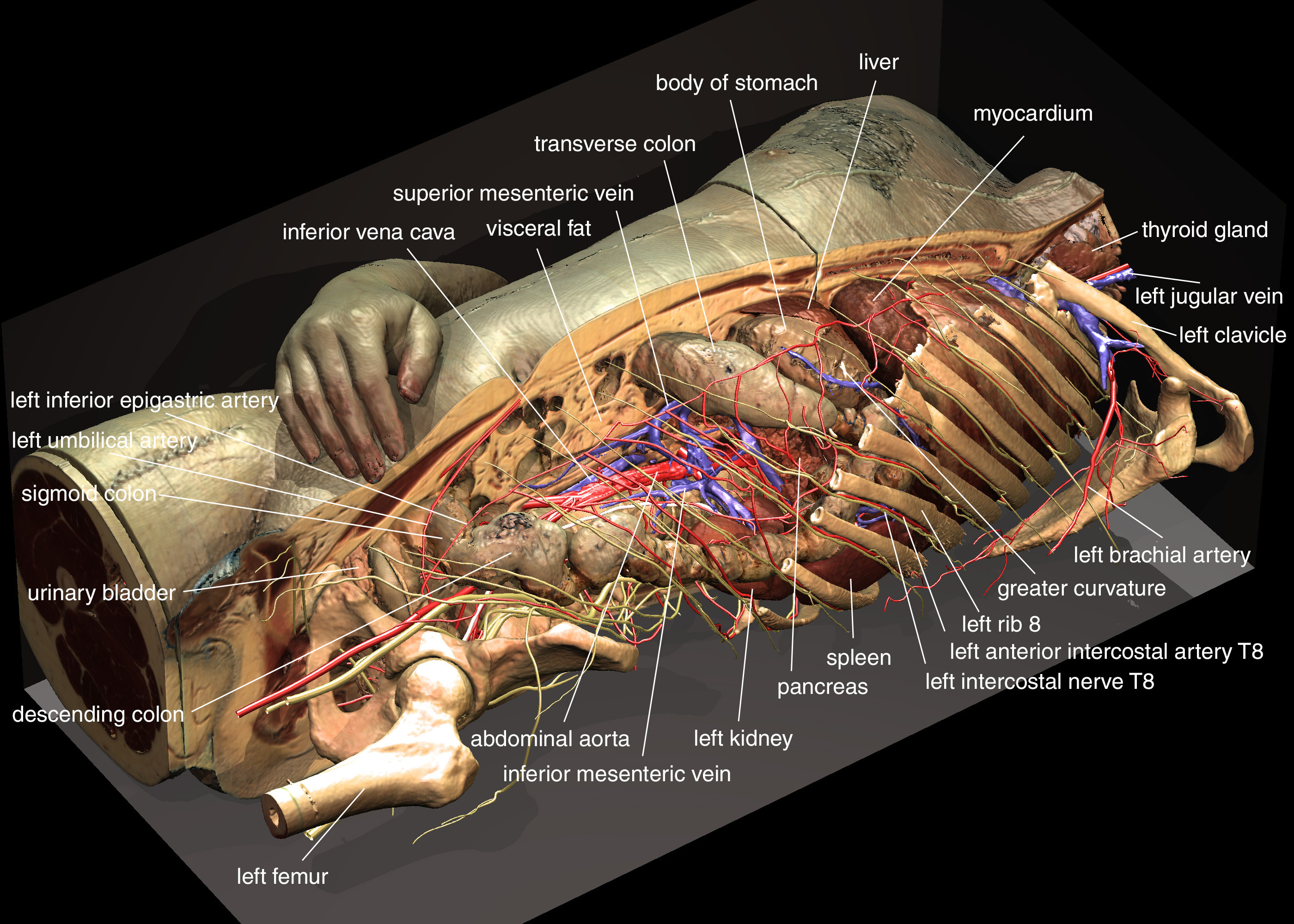
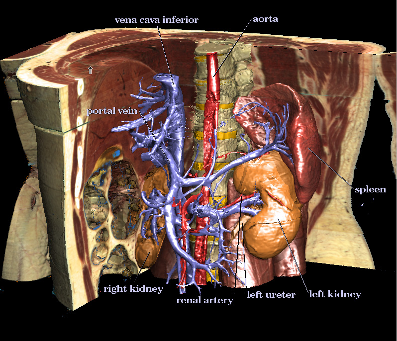
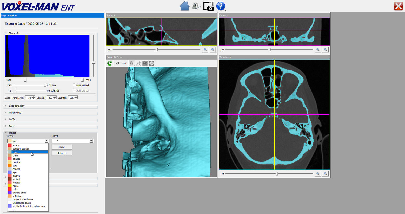
In 1995 the U.S. National Library of Medicine publishes the so called Visible-Human data set. It consists of 1878 photographic cross-sectional images of a corpse with the corresponding CT and MRI images. These data lend themselves for the creation of VOXEL-MAN 3D models. From these data the VOXEL-MAN team develops an interactive 3D atlas of the inner organs with unprecedented spatial resolution and fidelity. The team is supported by the expertise of Prof. Udo Schumacher (Institute of Anatomy and Experimental Morphology) and a committed crew of students helping with the elaborate work of segmentation (the assignment of voxels to organs). With the functionality known from the brain atlas 650 anatomical constituents can be examined. The graphic quality of the atlas published at Springer in 2001 is still (2019) unsurpassed. Now it is available for free download.
1998-2000 The Memorable Dissertation of Markus Urban
Markus Urban is a doctoral student at the Institute for Mathematics and Computer Science in Medicine. He is he is enthusiastic about medicine and a computer freak. He is given the task of creating a 3D atlas of the brain's vascular system from magnetic resonance imaging (MRI). He decides to do this using data from his own brain and undergoes lengthy imaging at Siemens in Erlangen. He does this with great expertise and tremendous energy. The result is a new type of interactive 3D atlas. Markus Urban dies in a traffic accident while riding his bicycle in December 2000 after a night shift in neuroradiology. The finished dissertation is on the table. The medical faculty awards him a doctorate posthumously.
2002 VOXEL-MAN receives Comenius Medal
The interactive anatomical atlases VOXEL-MAN 3D-Navigator: Internal Organs and VOXEL-MAN 3D-Navigator: Brain and Skull, published by Springer-Verlag, are awarded the 2002 Comenius Medal (now Comenius Edumedia Award).The Comenius Edumedia Award is the oldest European award for educational media.
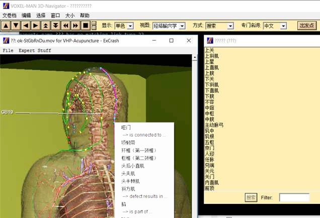
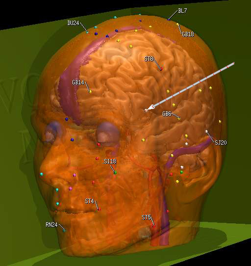
2002 A First 3D Acupuncture Atlas
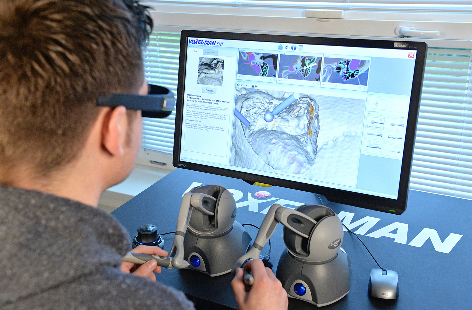
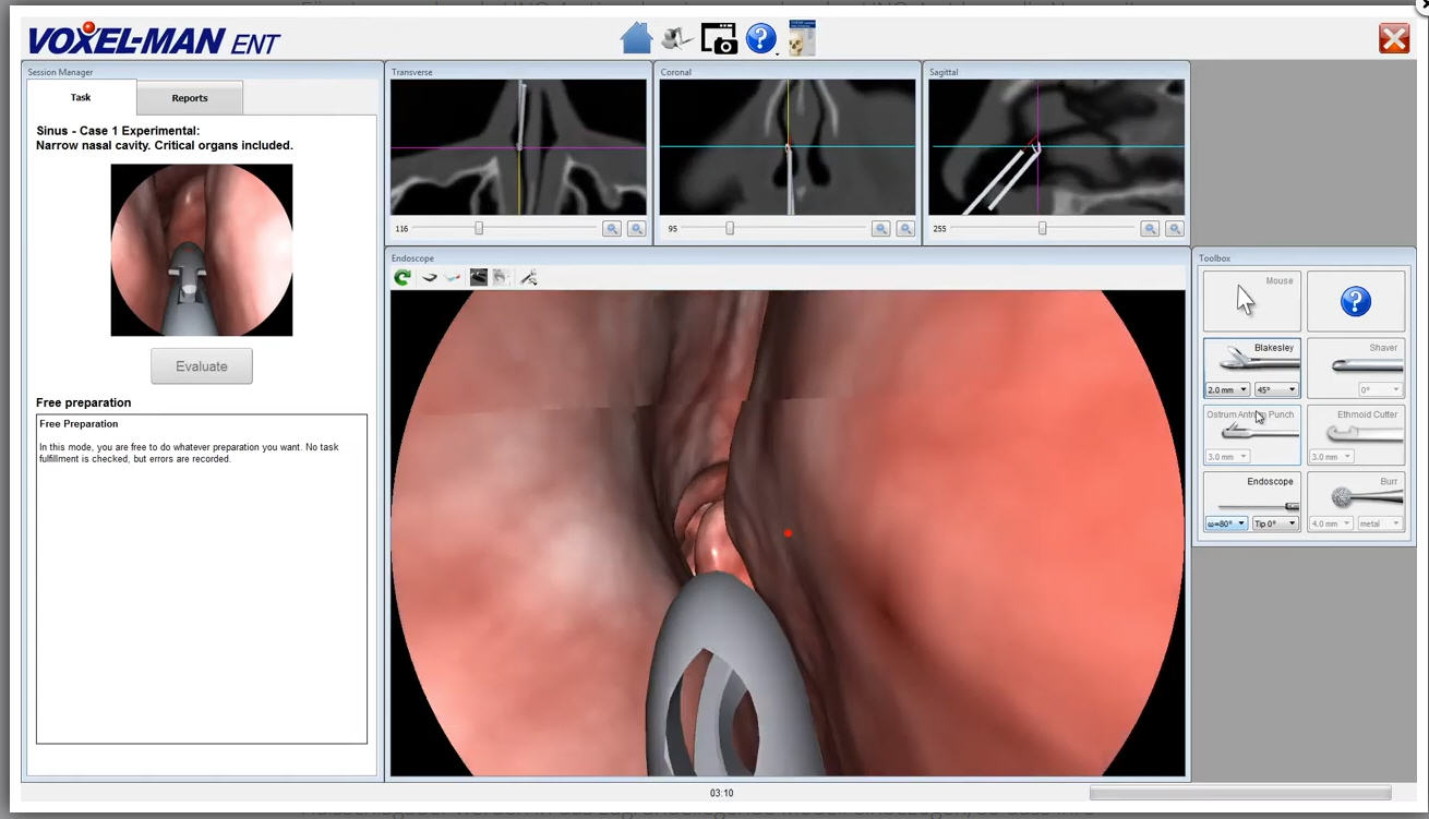
2003 The first Prototype of a Virtual Reality ENT Surgery Simulator
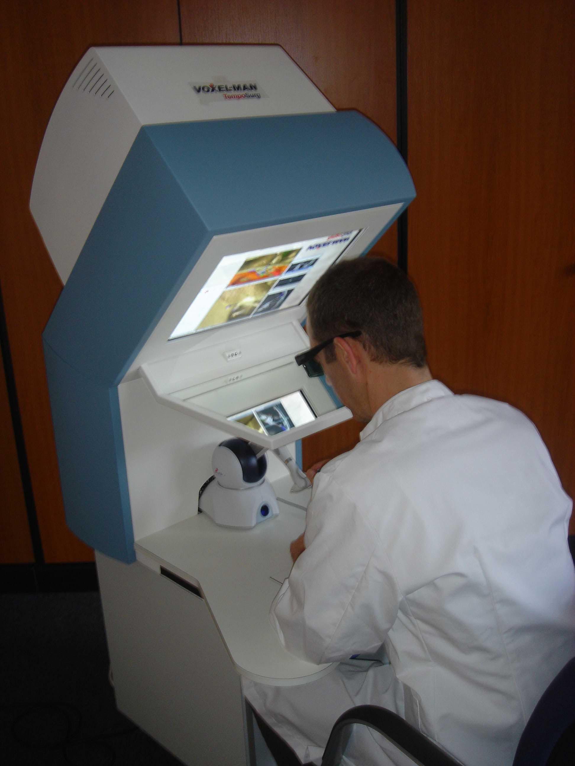
Gradually more surgeons find preoperative computer based 3D visualization useful, because it shows the anatomy as they find it at the intervention. The VOXEL-MAN team develops techniques for arbitrary cutting in 3D models. Using these, the simulation of risky interventions, such as those in the middle ear becomes possible. Via stereoscopic viewing and haptic force feedback device the surgeon gets the impression of acting on a real patient. Together with Dr. Rudolf Leuwer (ENT Department) the first virtual reality simulator prototype worldwide for middle ear surgery is presented.
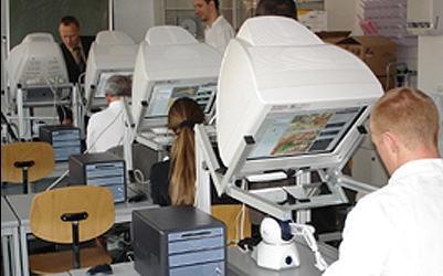
2005 The first Commercial Simulator for ENT Surgery
The company Spiggle und Theis Medizintechnik brings the system to market. Soon the system is sold to institutions worldwide for training in middle ear surgery.
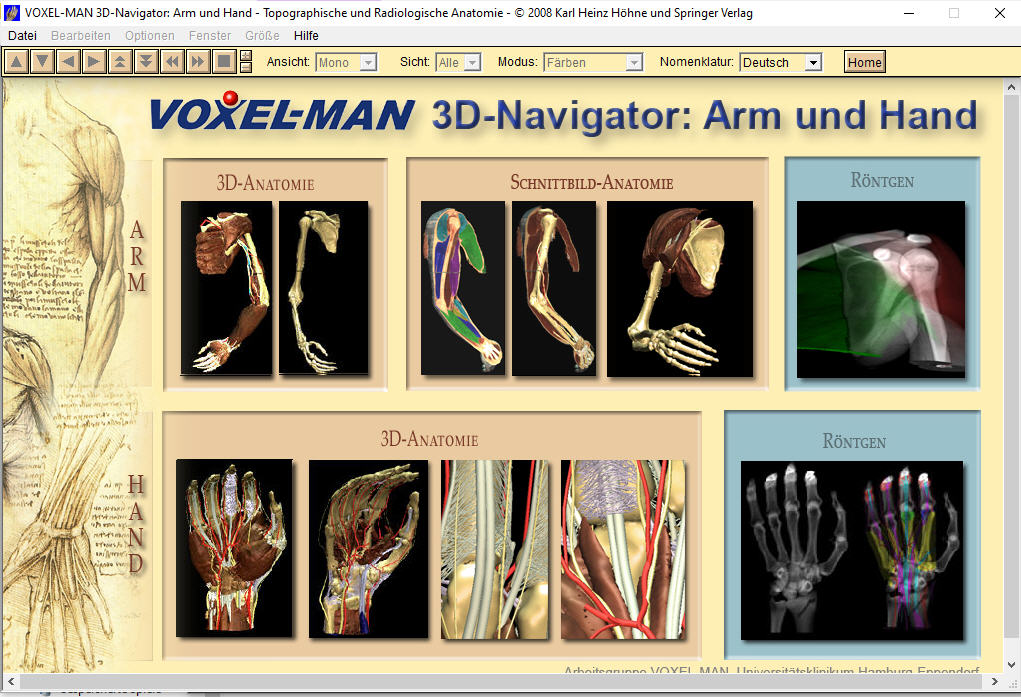
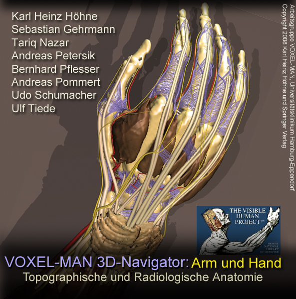
2008 VOXEL-MAN 3D Navigator Upper Limb
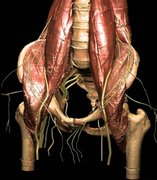
On the basis of the Visible-Human data set the 3D-Navigator Upper Limb, an interactive atlas of anatomy and radiology of the upper limb is developed and published at Springer.
2010 VOXEL-MAN becomes commercial activity of the University Medical Center
Led by Andreas Pommert and Ulf Tiede VOXEL-MAN becomes a commercial activity of the University Medical Center Hamburg-Eppendorf.
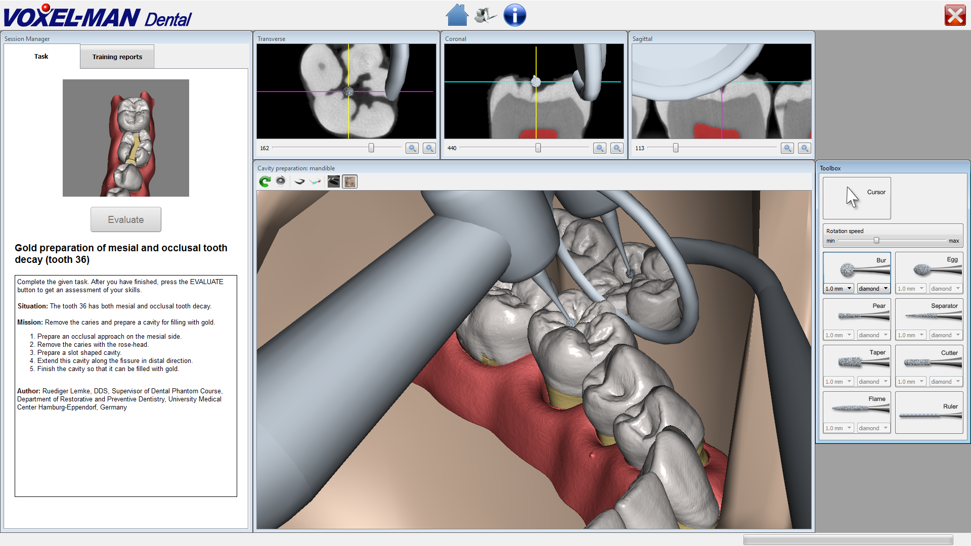
2011- today VOXEL-MAN as Leading Supplier for Simulators in ENT and Dentistry
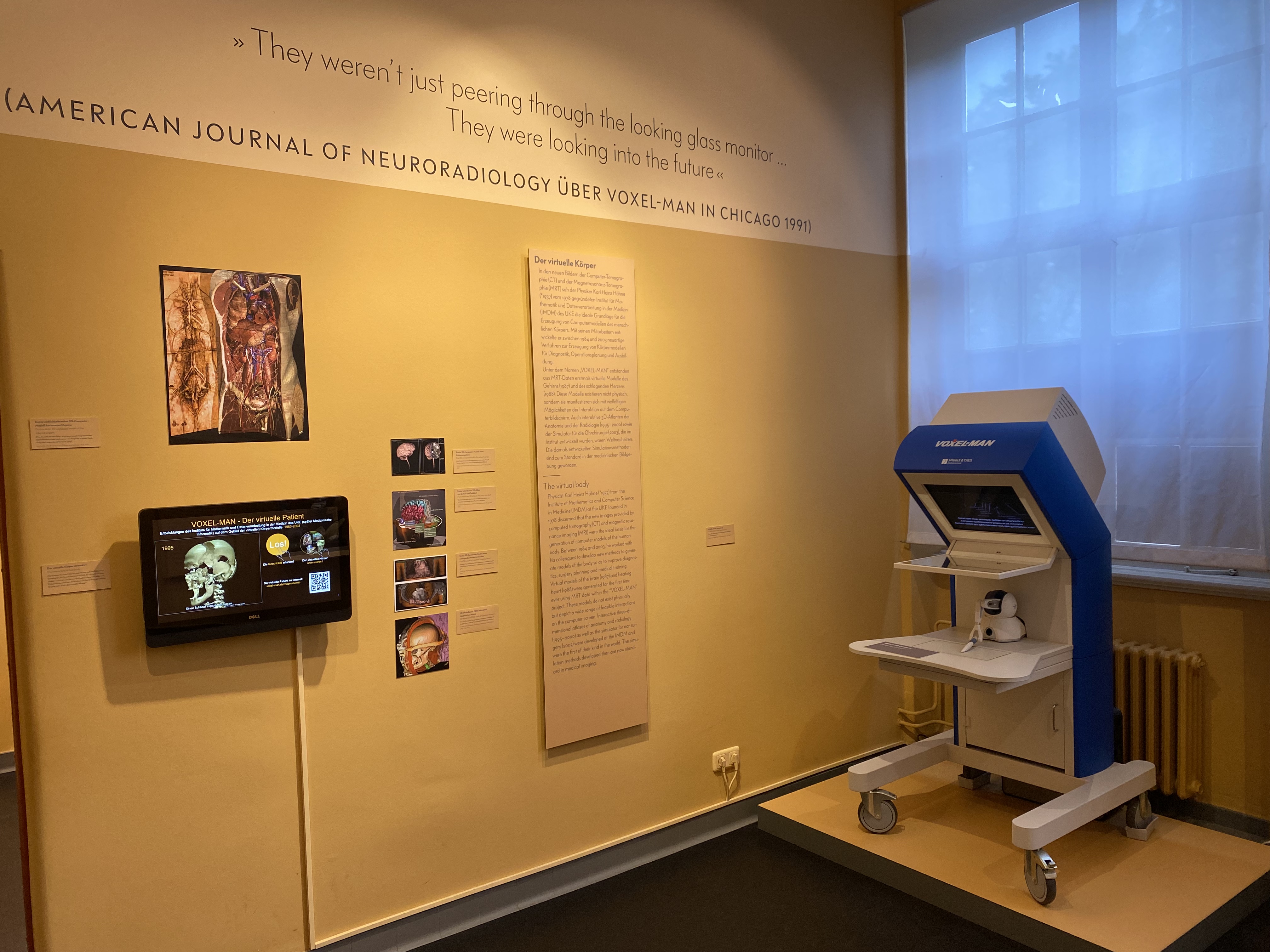
2013 VOXEL-MAN becomes part of the Hamburg Museum of Medical History
The early pioneering work of the project is included in the museum's permanent exhibition. 3D models of the human body can be examined on a touch screen, for example the first 3D model of a living patient's brain. Or you can see one of the first medical 3D prints. The process of simulating an intervention on the middle ear can be followed on the original version of the VOXEL-MAN ENT surgery simulator from 2005.
2020 MICCAI Enduring Impact Award
The achievements of the VOXEL-MAN project are acknowledged by the MICCAI Enduring Impact Award for Karl Heinz Höhne.
Methods developed for the VOXEL-MAN project also lead to new types of applications beyond medicine 1989-1997 'The Virtual Mummy'
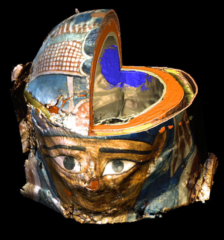
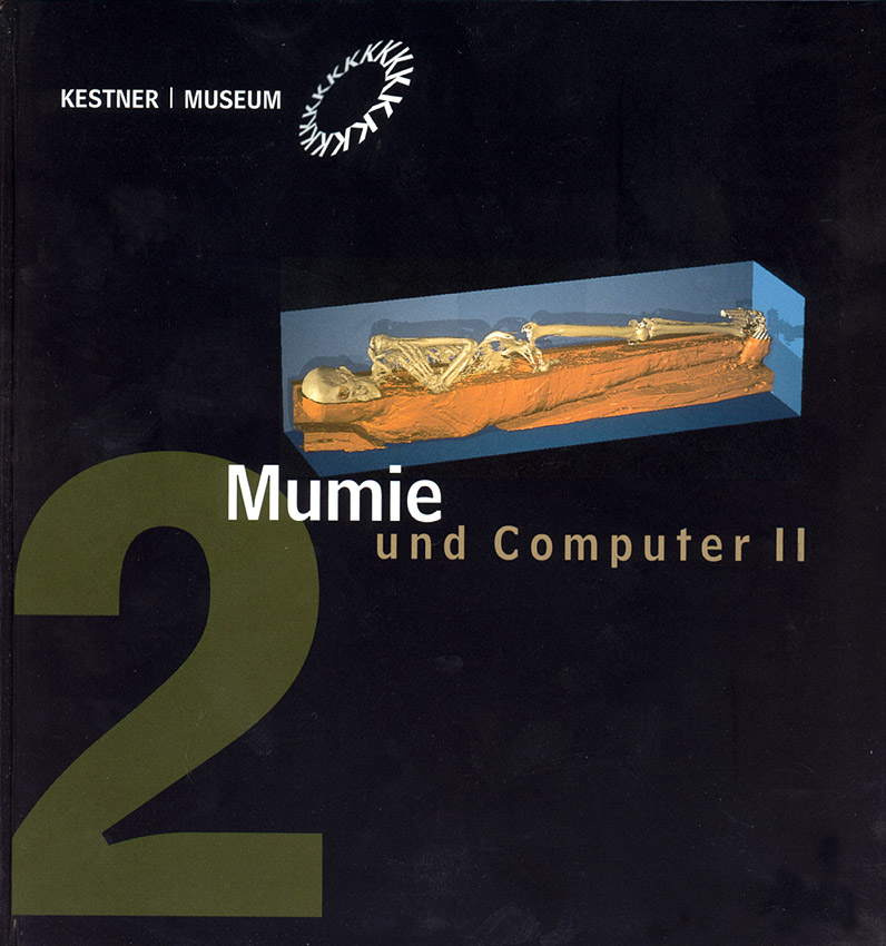
In 1989, at the occasion of a public lecture of Dr. Renate Germer (at that time with the Department of Egyptology of the University of Hamburg) the idea of transferring 3D modelling to mummy research is born. A 3D model of the head of a 30 year old woman passed away 2300 years ago is created from computer tomograms. It offers the possibility of a detailed investigation. As a "spin-off" from medical research a long term cooperation involving further mummies is established.1991/92 Reconstructions were shown at the exhibition Mumie + Computer at the Kestner Museum in Hannover. In 1997 the same data are reprocessed for the exhibition Mummies: Life after death in Ancient Egypt (Museum für Kunst und Gewerbe, Hamburg) and posted on the web as The Virtual Mummy. Around the year 2000 it is the most visited web page of the University Medical Center Hamburg-Eppendorf. Even the magazine Science* finds it worth mentioning under the title "Cool images". * Science 285, 491, 1999.
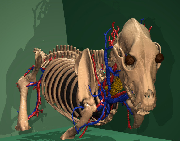
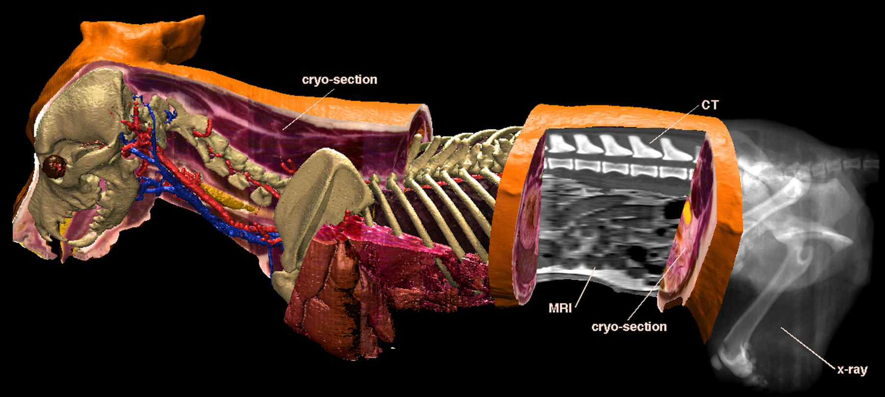
1999 'The Visible Dog' - 3D-Model of a Beagle
At the Institute of Vetereinary Anatomy of the Ludwig-Maximilians-University Munich Peter Böttcher acquires data of a beagle as
Using the VOXEL-MAN system he creates a three-dimensional atlas of the anatomy and radiology of a beagle.
|
|||||||||||||||||||||||||||||||||||||||||||||||||||||||||||||||||||||||||||||||||||||||||||||||||||||||||||||||||||||||||||||||||||||||||||||||||||||||||||||||||||||||||||||||||||||||||||||||||||||||||||||||||||||||||||||


| Special thanks go to the VOXEL-MAN group, particularly to Bastian Dittmar for continuously helping with the presentations for the Medical-Historic Museum. | |||||||||
 |
 |
 |
 |
 |
 |
 |
 |
 |
|
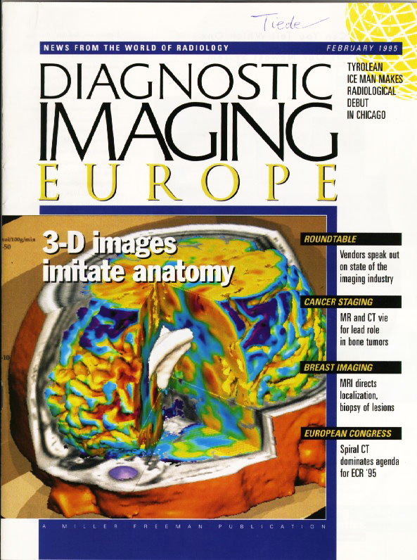
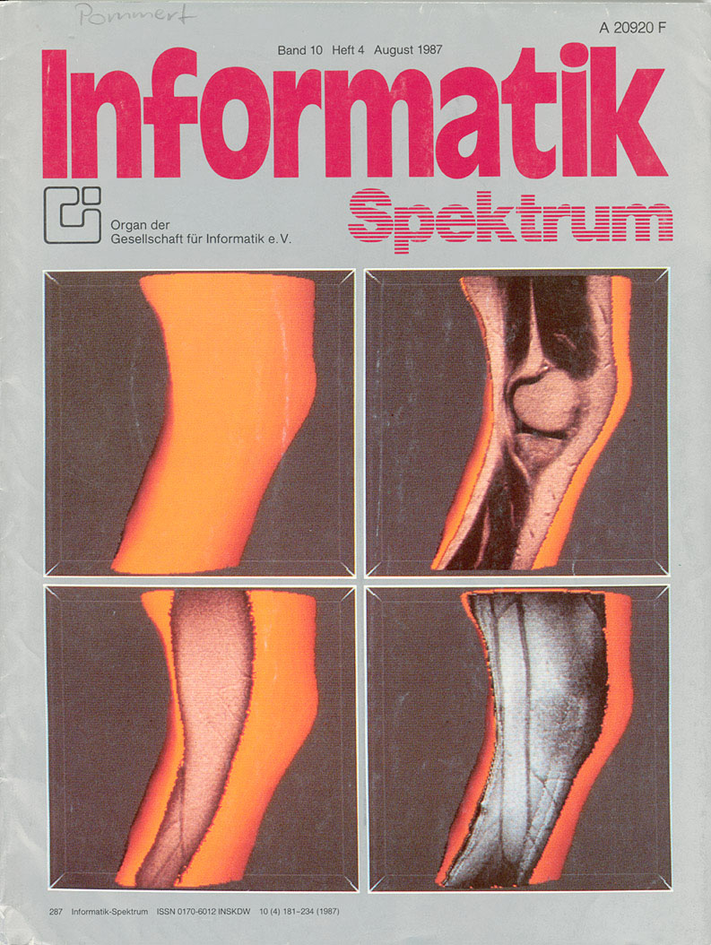
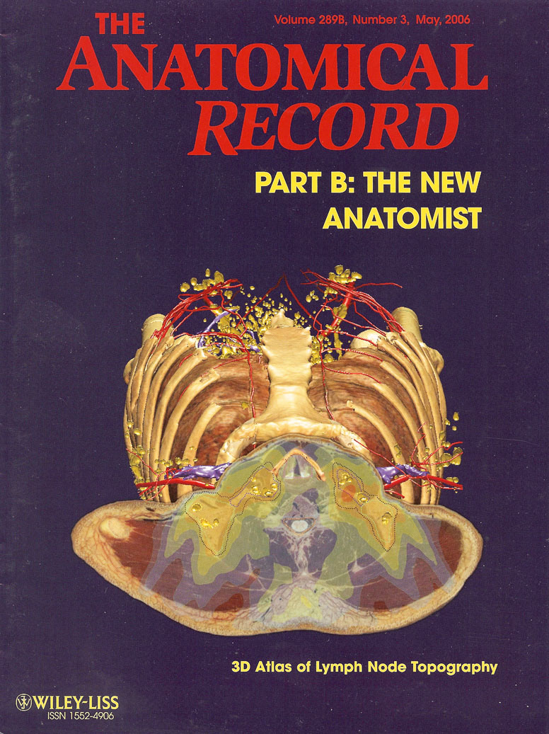
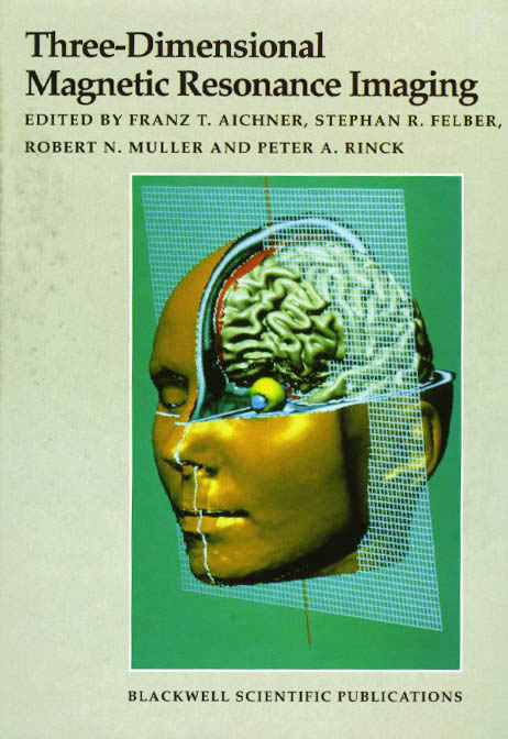
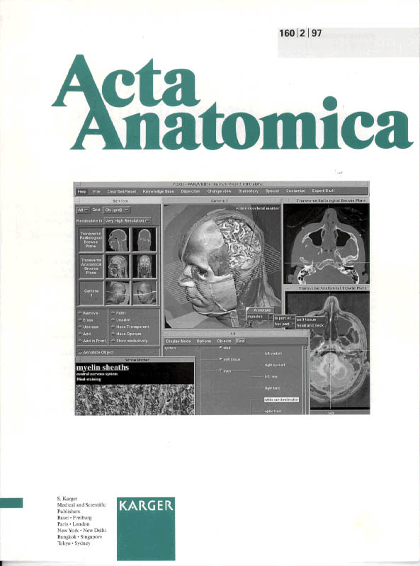
| VOXEL-MAN images as covers of journals and books | |||||||||
| mm The Virtual Patient |
|||||||||