 .
.Since 1984, the Institute for Mathematics and Data Processing at the University Hospital Hamburg-Eppendorf has been working on the development of three-dimensional representations from cross-sectional images from computer tomography (CT) and magnetic resonance imaging (MRI). One focus was the human brain. In 1987, the first realistic reconstruction of a patient's brain was achieved.
Video (16 Min.)
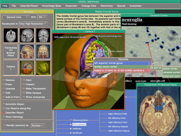
The first interactive 3D atlas of the anatomy and radiology of the human head followed in 1991 (VOXEL-MAN/brain and skull). It allows the anatomy to be rotated, disassembled, cut and queried at will, including its radiological appearance. Comment in the American Journal of Neuroradiology about exhibition visitors who worked with the atlas: ".................they were looking into the future". It turned out that the variety of possibilities overwhelmed a learner. Therefore, a simplified version for learners was developed, initially called VOXEL-MAN Junior, later called 3D Navigator/Brain and Skull. It consists of pre-calculated interactive scenes like the ones shown alongside.